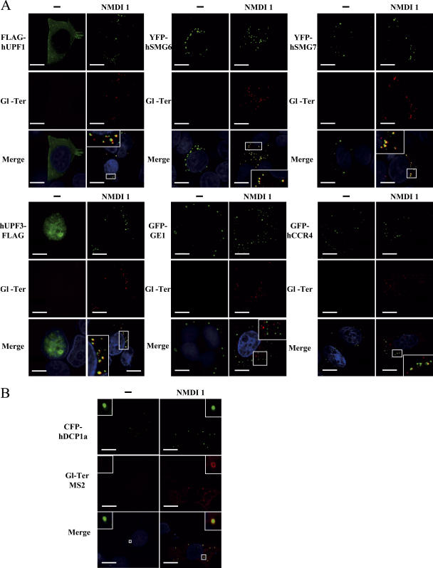
Figure 5.
PTC-containing mRNAs accumulate in P-Bodies under conditions of NMD inhibition. (A) HeLa cells were transfected with pmCMV-Gl Ter and an expression vector encoding a P-body component: pCI-neo-FLAG-hUpf1, pYFP-hSmg6, pYFP-hSmg7, pcDNA3-hUpf3-FLAG, pGFP-Ge1, or pGFP-hCcr4. 24 h after transfection, DMSO(−) or 5 μM NMDI 1 was added to the culture medium for 20 h. (B) U2OS cells were transfected with a pGl-Ter MS2 plasmid and a pCFP-hDcp1a expression vector. These cells were used for their tendency to highly express transfected genes, which is crucial for this approach. Cells were treated as described in A. Nuclei are shown in the merge by Hoechst staining in blue. The white squares are magnifications of cell areas. Bars, 10 μm.

