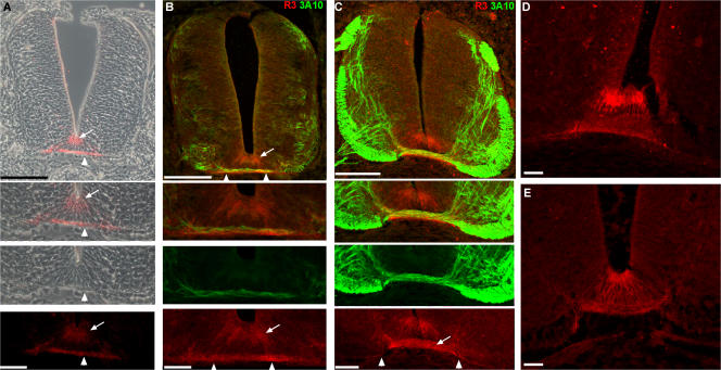
Figure 2.
F-spondin protein is deposited in the apical floor plate and the basement membrane, and binds to commissural axons. F-spondin protein expression at stage 21 (A and B), 26 (C and E), and 27 (D) chick embryos as revealed by the R3 antibody. F-spondin accumulates at the apical floor plate (arrow) and the basement membrane (arrowhead). (B) The pioneer commissural axons (3A10 staining) extend on the F-spondin (R3 staining) that is deposited in the basement membrane as they cross the midline at stage 21. (C) At stage 26, the immunoreactivity of F-spondin is evident on the crossing fibers of the commissural axons (arrow) and basement membrane that flanks the midline ventral pia (arrowheads). Inhibition of serine proteases by in ovo aprotinin injection reduces the deposition of F-spondin at the basement membrane (E) and yields homogenous staining along the surface of floor plate cells. Fluorescence and phase-contrast images in A were taken with microscope using a digital camera. Fluorescence images in B–E were taken with a confocal microscope. Bar, 40 μm.

