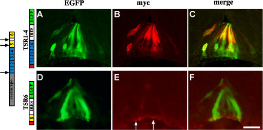
Figure 3.
Expression and protein localization of F-spondin domains at the floor plate. In all the experiments, enhancer III of HoxA-1 gene (Li and Lufkin, 2000) was used to drive expression of Cre recombinants (HoxA-1–Cre). A TSR1-4 conditional plasmid (loxP–STOP–loxP–TSR1-4–IRES–EGFP; A–C) and a TSR6 conditional plasmid (loxP–STOP–loxP–TSR6–IRES–EGFP; D–F) cloned in a pCAGG vector were electroporated into the spinal cord of stage 12–14 chick embryos. The F-spondin proteins were tagged with 4× myc epitope at their amino end. Cross-sections of stage 22–24 electroporated embryos were stained with antibodies to myc (B and E) and EGFP (A and D). TSR1-4 protein (B and C) labels the margins of cytoplasmic EGFP (A and C), reflecting its deposition along the membrane of the expressing cells. (E and F) TSR6 is deposited at the basement membrane that underlies the floor plate (arrows). (D) Note that the expressing cells, EGFP-positive cells, do not present the protein. The gray box represents the reelin/spondin domain of F-spondin. The blue boxes represent the nonadhesive TSR1-4 of F-spondin. The yellow boxes represent the adhesive TSR5 and 6 of F-spondin. The red box represents a cassette of 4× myc epitope. The black arrows point to the sites of cleavage of F-spondin. The white arrows point to the site of deposition of TSR6. Images were taken with a microscope using a digital camera. Bar, 25 μm.

