Figure 5.
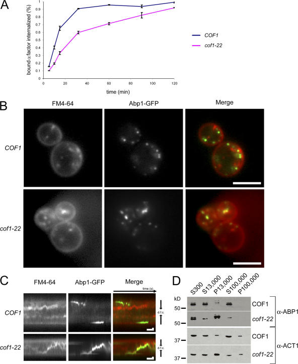
Cofilin defects do not block endocytic internalization but lead to Abp1 association with internal membranes. (A) Internalization of 35S-labeled α factor was measured at 25°C and displayed as percent internalized. Error bars represent the SD from two experiments performed in duplicate (four total measurements per time point). (B) Single medial focal plane image of cof1-22 cells expressing Abp1-GFP 5 min after the addition of FM4-64. Images were captured simultaneously. (C) Kymograph of a cof1-22 cell expressing Abp1-GFP 5 min after the addition of FM4-64. Kymographs represent fluorescent signals in the center of the cells over time. Bars in both kymographs are 5 s on the x axis and 500 nm on the y axis. dfc, distance from cortex. (D) Subcellular fractionation of wild-type and cof1-22 cell extracts using sequential centrifugation steps was used to generate low speed supernatant (S300), medium speed supernatant and pellet (S13,000 and P13,000), and high speed supernatant and pellet (S100,000 and P100,000) fractions. The subcellular distribution of Abp1p and Act1p was detected using immunoblotting with polyclonal anti-Abp1p and Act1p antibodies. Bars, 4 μm.

