Figure 6.
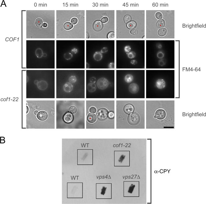
Defects in endocytic compartment morphology and secretion of the vacuolar hydrolase CPY in a cofilin mutant. (A) Representative medial focal plane images of endocytic internalization of the lipophilic dye FM4-64 in wild-type and cof1-22 mutant cells. Vacuoles identified by brightfield microscopy are marked with red asterisks. (B) Colony blot assay for CPY secretion in wild-type cells and cof1-22, vps4Δ, and vps27Δ mutants grown at 25°C. Bar, 4 μm.

