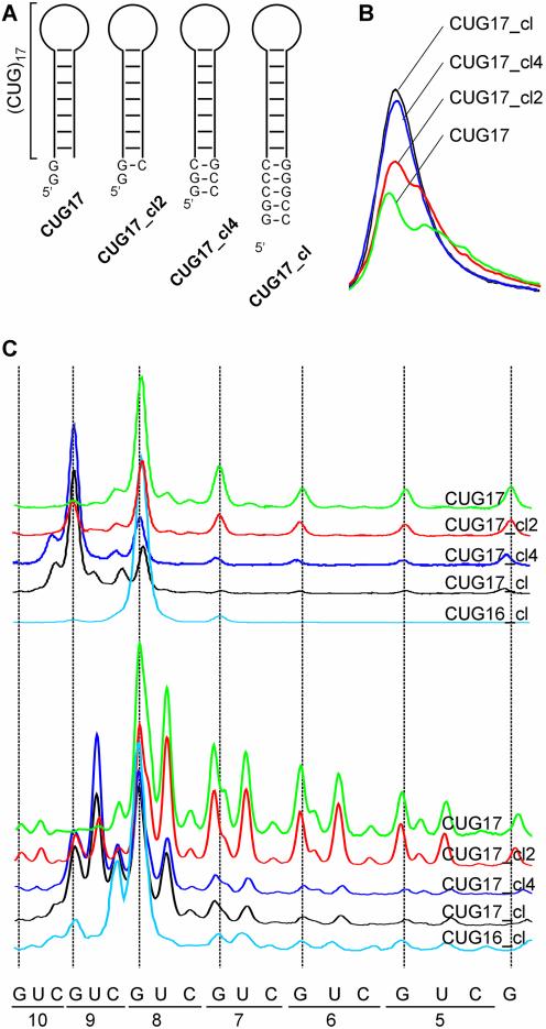
Figure 2.
Structure analysis of (CUG)n repeat transcripts having the GC-clamp. (A) Schematic secondary structures of four transcripts, CUG17, CUG17_cl2, CUG17_cl4 and CUG17_cl, used in this analysis. (B) Qualitative comparison of 5′-end labeled transcripts shown in (A) containing a different number of base pairs forming a GC-clamp, obtained from PhosphorImaging. All transcripts were analyzed in the same polyacrylamide gel in non-denaturing conditions. (C) Cleavage patterns obtained for 5′-end labeled transcripts described above using T1 ribonuclease (upper panel) and S1 nuclease (lower panel). Results obtained for the CUG16_cl transcript were added to show its structural similarity to non-clamped transcripts.

