Figure 3.
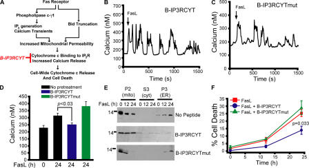
Cytochrome c binding to IP3R contributes to a late phase of calcium elevation in response to Fas ligand. (A) Schematic diagram depicting the predicted steps leading to calcium mobilization during FasL stimulation. B-IP3RCYT (in red) blocks cytochrome c binding to IP3R (Boehning et al., 2005). (B and C) Calcium responses in cells pretreated with 400 nM B-IP3RCYT or B-IP3RCYTmut, which does not bind cytochrome c. (D) Cytoplasmic calcium concentration in cells treated for 24 h with FasL in the presence or absence of 400 nM of the indicated peptide. (E) Subcellular fractionation of cells stimulated with FasL for 12 or 24 h and pretreated with vehicle or 400 nM peptide. (F) Cell death curve of control or peptide-pretreated cells. Data is presented as the mean ± the SEM.

