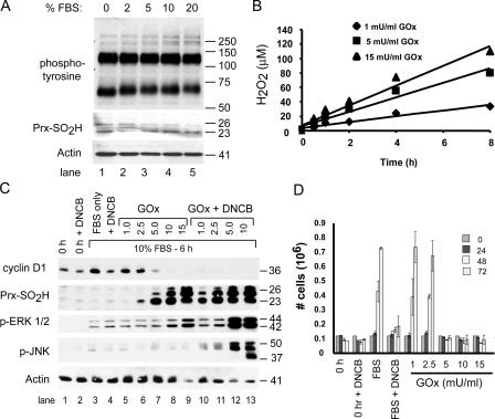
Figure 2.
Hyperoxidation of Prxs in serum-stimulated cells correlates with inhibition of cell proliferation. (A) C10 cells synchronized by serum deprivation were stimulated with medium containing the indicated concentration of FBS for 15 min, and levels of phosphotyrosine and Prx-SO2H were assessed by immunoblotting. Actin was used as a loading control. (B) For generating fluxes of H2O2, GOx was added to DME with 10% FBS at the indicated concentration, and the amount of H2O2 in medium was measured as a function of time. (C) Serum-starved mouse lung epithelial (C10) cells (time 0) were stimulated with DME containing 10% FBS and the indicated concentrations of GOx (mU/ml), with or without 5 μM DNCB, for 6 h. Cell extracts were examined for expression of the indicated proteins by immunoblotting. PrxI and -II comigrate under reducing conditions on SDS gels; based on molecular mass, the band at 26 kD is mitochondrial PrxIII. (D) The indicated concentrations of GOx were added to culture medium during serum stimulation, and replicate C10 cultures were counted over a 3-d period to assess cell proliferation. Control cultures (time 0) were maintained in DME with 0.5% FBS for the duration of the experiment or stimulated with 10% FBS alone. Error bars indicate mean ± SD.

