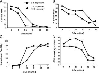
Figure 4.
Prx oxidation occurs before depletion of cellular GSH. Using the strip–reprobe immunoblotting method, densitometry was used to quantify the signals for the fraction of PrxI (A) or PrxII (B) that was not hyperoxidized as a function of GOx concentration after 3 h of exposure to the indicated concentration of GOx (squares) or after a 3-h recovery period (diamonds). (C) Total Prx-SO2H levels are expressed as a percentage of the maximal signal obtained with 15 mU/ml GOx, which caused quantitative hyperoxidation of PrxI and -II. (D) Reduced GSH levels were measured in extracts from C10 cells treated with GOx and allowed to recover in the same fashion. Error bars indicate mean ± SD.

