Figure 6.
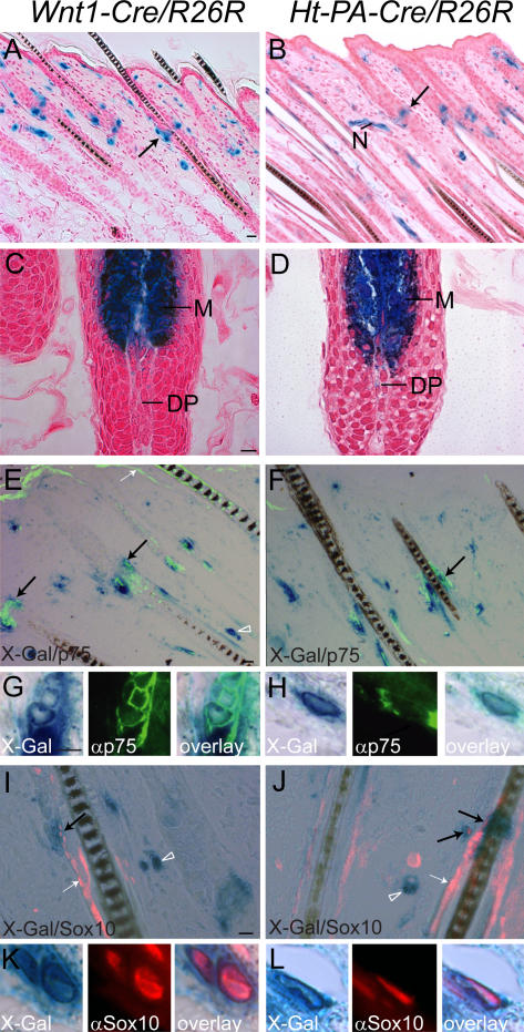
Localization of neural crest–derived and p75/Sox10-positive cells in murine back skin. Back skin from Wnt1-Cre/R26R (A, C, E, G, I, and K) and Ht-PA-Cre/R26R (B, D, F, H, J, and L) mice with pelage follicles in anagen phase was stained with X-gal solution. In both transgenic mouse models, positive cells were localized in nerves (N), in the hair follicle bulge region below the sebaceous gland (arrows; A and B), and in the bulb containing melanocytes (M; C and D). E, F, I, and J show pictures of back skin sections stained with X-gal and either antibodies against p75 or against Sox10 visualized by immunofluorescence. Enlarged areas show X-gal–positive cells in the bulge area coexpressing p75 (G and H) and Sox10 (K and L). The panels to the right represent overlays of X-gal and marker stainings. Note that although p75/X-gal– and Sox10/X-gal–positive cells in the bulge area point to the existence of NCSC-like cells in this location, p75 and Sox10 are not specific for neural crest derivatives in the skin and also label X-gal–negative structures (white arrows). Moreover, not all neural crest–derived cells coexpress p75 and Sox10 (arrowheads). The dermal papilla (DP) was X-gal negative in both mouse lines. Bars: (A, C, E, and I) 20 μm; (G) 10 μm.

