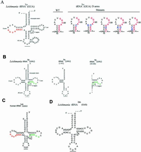
Figure 1.
Structures of tRNAs and their derivatives. (A) Left, Leishmania tRNATyr(GUA). The conserved D arm signal is shown in red. Right, D arm mini-helices, with mutations in blue. (B) Left, Leishmania tRNAIle(UAU); the conserved V-T region signal is shown in green. Right, truncated versions of tRNAIle[1–41] and tRNAIle[42–66]. (C) Human cytoplasmic tRNALys(UUU) with conserved D arm (red) and V-T (green) motifs. (D) tRNAGln(CUG).

