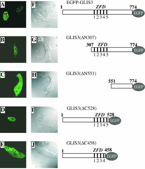
Figure 6.
GLIS3 localizes primarily to the nucleus. Plasmids pEGFP-GLIS3 (A and F), pEGFP-GLIS3(ΔN307) (B and G), pEGFP-GLIS3(ΔN551) (C and H), pEGFP-GLIS3(ΔC528) (D and I) or pEGFP-GLIS3(ΔC458) (E and J) were transfected into CV-1 cells and after 30 h the cellular localization of EGFP–GLIS3 fusion proteins examined by fluorescence confocal microscopy as described in Materials and Methods (A–E). EGFP was equally divided between cytoplasm and nucleus (not shown). 1–5 indicate the five zinc finger motifs. (F)–(J) Confocal images.

