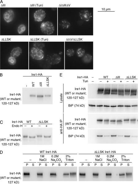
Figure 6.
Cluster formation, localization, cellular expression, or BiP dissociation is not significantly altered by either the ΔIII or the ΔLLSK mutation. (A) KMY1516 (ire1Δ) cells carrying the indicated mutant version of pRS423-IRE1-HA were subjected to anti-HA immunofluorescent staining. Cells were observed by a conventional fluorescent microscope, and exposure times for image acquisition were 1 s for all panels. (B and C) Lysates from KMY1015 (ire1Δ) cells carrying pRS315-IRE1-HA (wild type [WT] or mutant) were analyzed by anti-HA Western blotting. (C) Cell lysate was treated with Endo H where indicated. (D) Detergent-free crude cell lysate containing 0.7 M sorbitol was prepared from KMY1516 cells carrying pRS423-IRE1-HA (WT or ΔLLSK) and incubated with the indicated reagents (final concentration) on ice for 10 min. The samples were then centrifuged at 100,000 g for 30 min, and pellet (P) and supernatant (S) fractions prepared from the same amount of samples were analyzed by anti-HA Western blotting. (E) Lysates from KMY1516 cells carrying pRS423-IRE1-HA (WT or mutant) were subjected to anti-HA immunoprecipitation, and the lysates and the IPs were analyzed by anti-HA or anti-BiP Western blotting to detect the indicated proteins. When indicated in A and E, Tun was added at 2-μg/ml final concentration into cultures 1 h before harvest. For vector control (−), cells carried an empty vector, pRS315 (B) or pRS423 (E).

