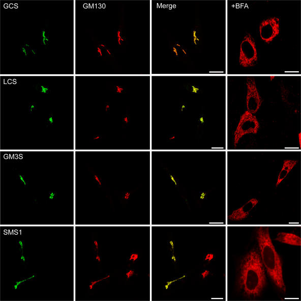
Figure 1.
Cellular localization of enzymes in Golgi sphingolipid synthesis by confocal fluorescence microscopy. HeLa cells were transiently transfected with the epitope-tagged enzymes (Table I). After 18 h, cells were fixed, permeabilized, and labeled with rabbit antibodies against the HA or V5 epitopes and with mouse antibody against GM130, a cis-Golgi matrix protein. Cells were counterstained with FITC-labeled anti–rabbit (left) and Texas red–labeled anti–mouse (GM130) antisera. Overlapping distributions appear as yellow in the merged images. After brefeldin A (BFA) treatment for 0.5 h, the glycosyltransferases were labeled with the specific antibodies followed by Texas red–labeled secondary antibodies. BFA fuses the Golgi stack to the ER. GCS, GlcCer synthase; LCS, LacCer synthase; GM3S, GM3 synthase; SMS1, SM synthase 1. For GCS, the transfected cells displayed 6.4× the activity of wild-type cells. Bars, 10 μm.

