Figure 5.
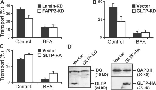
Effect of the expression levels of FAPP2 and GLTP on GSL transport to the cell surface. (A) MEB4 cells stably expressing RNAi plasmids against FAPP2 or lamin were preincubated with or without 1 μg/ml BFA for 0.5 h and labeled with [14C]palmitic acid for 1.5 h. [14C]GSLs were extracted from the cell surface during an additional 45-min incubation with 1.5 mg/ml GLTP in the medium. The lipids in the cells and media combined with the washes were analyzed and quantified. Transport is expressed as the percentage of GlcCer recovered in the medium. (B and C) D6P2T cells stably expressing RNAi plasmids against GLTP or empty vector (B) or stably transfected with GLTP (C) were treated with BFA and labeled with [14C]galactose, and transport of [14C]GlcCer to the cell surface was measured as in A. Error bars represent SEM. (D) Western blot analysis from D6P2T clones expressing either RNAi plasmids (B) against GLTP and empty vector as control or pCDNA3.1 plasmids with GLTP-HA (C) using a rabbit antiserum against mouse GLTP. A background band at ∼40 kD (BG) and a blot against glyceraldehyde-3 phosphate dehydrogenase (GAPDH) were used as loading controls.

