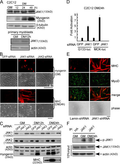
Figure 1.
JAK1 knockdown accelerates myogenic differentiation. (A) WCEs from either C2C12 or primary myoblasts were subjected to Western blotting for various markers. (B and C) C2C12 cells were transfected with various siRNAs as indicated. (B) Cells were fixed at different time points as indicated and subjected to immunostaining for either myogenin or MHC. Phase-contrast images of the same field are also shown. (C) WCEs were prepared, and 20 μg WCE was subjected to immunoblotting. (D) Duplicate C2C12 cells were transfected with siRNAs and the luciferase reporter constructs as indicated. 24 h after transfection, cells were switched to DM for another 24 h. WCEs were prepared and subjected to luciferase assays. Fold activation was calculated as the ratio of the luciferase activity of the JAK1-siRNA–transfected cells over that of the GFP-siRNA–transfected cells. The results are presented as mean ± SD (error bars). (E) Primary myoblasts were infected with adenoviruses expressing either JAK1-shRNA or lamin-shRNA. 3 d after infection, cells were fixed and subjected to double immunostaining for MyoD and MHC. (F) WCEs were prepared from C2C12 cells grown in GM or DM for 24 h. The endogenous JAK1 was immunoprecipitated (IP) from 1 mg WCE and subjected to in vitro kinase assays. The HA antibody was used as a negative control. Bars, 100 μm.

