Figure 4.
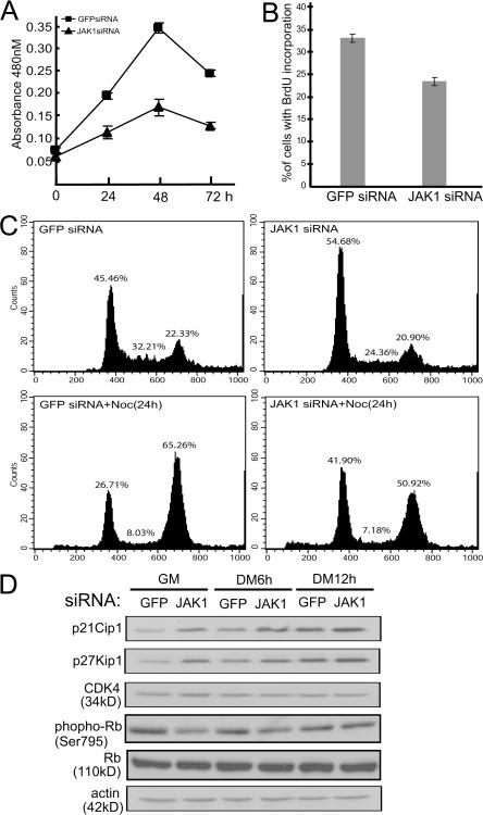
JAK1-siRNA inhibits myoblast proliferation. C2C12 cells were transfected with either GFP-siRNA or JAK1-siRNA as indicated. (A) WST-1 reagent was added to cells at different times as indicated, and absorbance at 480 nm was measured by a plate reader. The experiment was performed in triplicate, and the results are presented as mean ± SD (error bars). (B and C) 30 h after siRNA transfection, 10 μM BrdU was added for 1 h (B). Cells were fixed and subjected to immunostaining using antibodies against BrdU. Nuclei were counterstained with DAPI, and images were taken and analyzed by fluorescent microscopy. The percentage of cells positive for BrdU staining was calculated based on cells from five randomly chosen fields (10× magnifications). The results are presented as mean ± SD. (C, top) Cells were trypsinized, fixed, and subjected to FACS analysis. (bottom) Cells were treated with nocodazole for 24 h followed by FACS analysis. (D) WCE was prepared at different times as indicated, and 30 μg WCE was subjected to immunoblotting analysis.

