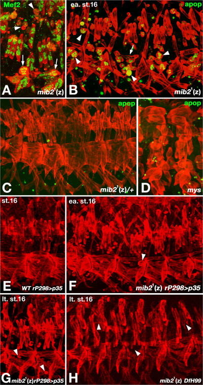
Figure 4.
mib2 activity is required for preventing apoptosis of somatic muscles. In all panels, muscles were labeled with anti-Tropomyosin (red). (A) Zygotically (z) mib21 homozygous mutant embryo at stage 16 with disappearance of Mef2-labeled nuclei in shrunken syncytia (arrowheads). Arrows indicate Mef2-positive muscles at an earlier stage of detachment. (B) Embryo as in A but labeled with TUNEL (green), showing strong apoptotic signals in the detached and shrinking muscle syncytia (arrowheads). Arrow indicates detaching muscle with weak apoptotic signals. (C) Stage 16 mib21 heterozygous control embryo without any apoptotic signals in somatic muscles. (D) Stage 16 myospheroid (mys) mutant embryo with an absence of apoptotic signals in detached muscles which are not shrunken. (E) Stage 16 control embryos with p35 overexpression via rP298-Gal4 exhibit normal somatic muscle morphology. (F) Early stage 16 mib21 (z) embryo with forced expression of p35 via rP298-Gal4, showing only a few detached muscles (arrowheads). (G) Late stage 16 embryo as in (F) with only a few detached or missing muscles (arrowheads). (H) Late stage 16 embryo homozygous for mib21 and Df(3R)H99. Most somatic muscles appear normal although a few have aberrant shapes or are missing (arrowheads).

