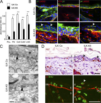
Figure 3.
ILK ablation impairs keratinocyte adhesion, integrin expression, and BM integrity and alters proliferation in vivo. (A) Cell adhesion of ILK-K5 keratinocytes from 4-d-old mice on FN, Col I, Col IV, and LM332 is significantly reduced compared with control keratinocytes. Adhesion to poly-l-lysine (PLL) is not different (mean + SD of three independent experiments; ***, P < 0.001). (B) Integrin expression in epidermis from 2-wk-old mice. In control skin, β1 and β4 integrins are expressed in basal keratinocytes, whereas in ILK-K5 skin both integrins are also found on suprabasal keratinocytes (arrowheads). In ILK-K5 mice, β4 integrin shows discontinuous staining on basal keratinocytes, LM332 diffuses into the upper dermis (asterisks), and β1 integrin–expressing suprabasal cells retain K14 expression. Bars, 25 μm. (C) Electron micrographs of back skin sections of 2-wk-old control and ILK-K5 mice. Control skin exhibits a continuous lamina densa (asterisks) and hemidesmosomes (arrowhead), whereas ILK-K5 skin shows a discontinuous lamina densa, which is preserved at hemidesmosomes (arrowhead) but largely absent in between. Bar, 0.25 μm. (D) Ki67 staining revealed the presence of proliferating cells in ILK-K5 suprabasal layers. Suprabasal BrdU+ cells express β4 integrin. Bars, 25 μm.

