Figure 7.
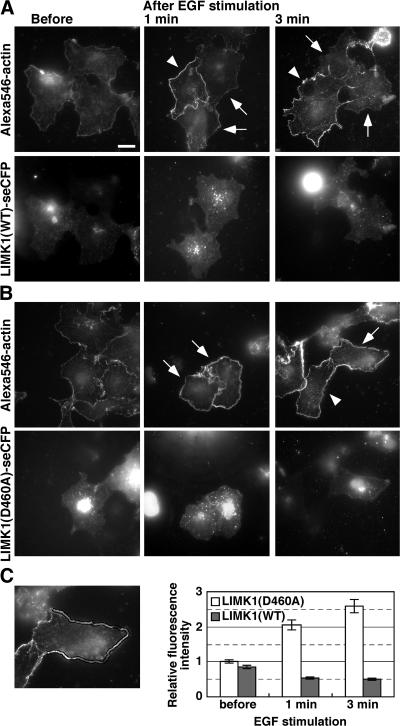
Cofilin inactivation suppresses EGF-induced barbed end formation in the cell periphery. (A and B) COS7 cells transfected with LIMK1(WT)-SECFP (A) or LIMK1(D460A)-SECFP (B) were left unstimulated or stimulated with EGF for 1–3 min. Cells were permeabilized with nucleation buffer containing 0.025% saponin and 0.45 μM Alexa546-actin monomers, washed three times, and fixed to analyze Alexa546-actin (top) and LIMK1-SECFP fluorescence (bottom) using fluorescence microscopy. Arrows indicate LIMK1(WT)- or LIMK1(D460A)-expressing cells, and arrowheads indicate nonexpressing cells, assessed by SECFP fluorescence. Bar, 20 μm. (C) Quantitative analysis of Alexa546-actin fluorescence in the cell periphery. Incorporation of Alexa546-actin into the cell periphery was measured as the mean fluorescence intensity in a region 2 μm from the cell edge, using ImageJ. The left image shows the region measured. Data are means ± SEM of 38–42 cells from three independent experiments, with the mean fluorescence in unstimulated LIMK1(D460A)-expressing cells normalized to 1.0.

