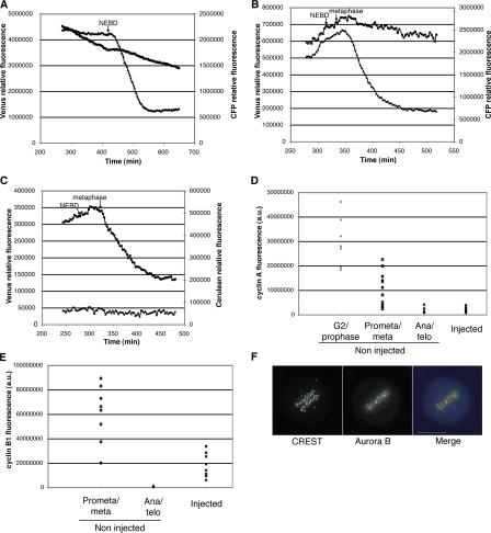
Figure 2.
The timing of degradation of cyclins A and B1 and securin is not affected by the presence of Emi1. (A) Cyclin A–CFP: HeLa cells were coinjected in G2 phase with Venus-Emi1 D144A and cyclin A–CFP and followed by time-lapse microscopy. The fluorescence levels of both proteins were measured and plotted over time. NEBD was determined from the DIC images. This result is representative of four cells from two experiments with Venus-Emi1 D144A, 14 cells from three experiments with Venus-Emi1 G146V, and eight cells from five experiments with Venus-Emi1 S145A/S149A. The apparent decrease of Venus-Emi1 D144A fluorescence is the result of photobleaching. (B) Cyclin B1–CFP: HeLa cells were coinjected in G2 phase with Venus-Emi1 D144A and cyclin B1–CFP and analyzed as in A. This result is representative of 15 cells in six experiments with Venus-Emi1 D144A and six cells in three experiments with Venus-Emi1 G146V. (C) Securin- Cerulean: HeLa cells were coinjected in G2 phase with Venus-Emi1 D144A and securin-Cerulean followed by time-lapse fluorescence microscopy, and the fluorescence levels were measured and analyzed as in A and B. This result is representative of seven cells in three experiments with Venus-Emi1 D144A. Note that in these experiments, lower amounts of Venus-Emi1 D144A were injected. (D) Cyclin A: HeLa cells injected with Venus-Emi1 D144A in G2 phase were followed by time-lapse microscopy and fixed after they had delayed in metaphase. Cells were stained for cyclin A, and the levels were quantified in the injected arrested cells and in control noninjected cells at different stages. The quantification shown is of PFA-fixed cells, and similar results were obtained after methanol/acetone fixation. Similar results were obtained with two different cyclin A antibodies. Results are representative of four experiments. (E) Cyclin B1 levels analyzed on cells injected, filmed, and stained as in D. The quantification shown is of cells fixed in methanol/acetone, and similar results were obtained after PFA fixation. Results are representative of five experiments. (F) HeLa cells that delay in mitosis after Venus-Emi1 D144A injection (blue in the merge) were fixed and stained with CREST serum (red in the merge) and aurora B antibodies (green in the merge). Representative of 11 cells analyzed. Bar, 15 μm.

