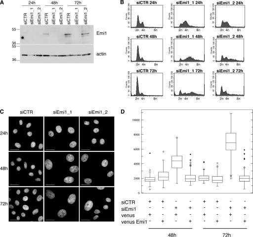
Figure 5.
Emi1 is required to prevent rereplication. (A) Asynchronous HeLa cells were transfected with control or two different Emi1 siRNAs, and Emi1 levels were analyzed by Western blotting 24, 48, and 72 h after transfection. Actin is shown as a loading control. Results are representative of four experiments. (B) DNA content of cells transfected as in A and analyzed by flow cytometry after staining with propidium iodide. Results are representative of four experiments. (C) Cells transfected as in A were stained with Hoechst to visualize the nuclei. Bars, 30 μm. (D) Cells cotransfected with control and Emi1 siRNAs plus expression vectors encoding Venus (control) or Venus-Emi1 (as indicated) were fixed 48 and 72 h after transfection, and DNA was stained with Hoechst. The nuclear size (in pixels on the y axis) was measured using ImageJ software. The small bars in the dot plot show the minimum and maximum values, and the box shows the first and third quartiles. The bar in the box is the median value. Outliers (closed circles) and suspected outliers (open circles) as determined by statistical analysis are shown. Results are representative of two independent experiments. More than 100 nuclei were measured for each sample.

