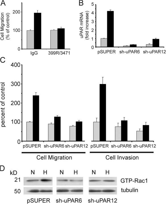
Figure 4.
uPAR is necessary for hypoxia-promoted cell migration, invasion, and Rac1 activation. (A) MDA-MB-468 cells were pretreated with 25 μg/ml uPA-specific antibody 3471 and 25 μg/ml uPAR-specific antibody 399R or with 50 μg/ml control IgG. Cells were allowed to migrate in Transwell chambers for 24 h in 21% O2 (gray bars) or 1.0% O2 (black bars). Cell migration is expressed as a percentage of that observed with control IgG in normoxia (mean ± SEM; n = 6). (B) MDA-MB-468 cells expressing empty vector (pSUPER), sh-uPAR6 cells, and sh-uPAR12 cells were cultured for 24 h in 21% O2 (gray bars) or 1.0% O2 (black bars). uPAR mRNA was determined by qPCR. Results are normalized against HPRT-1 and compared with the level observed in the pSUPER cells in normoxia (mean ± SEM; n = 3). (C) pSUPER, sh-uPAR6, and sh-uPAR12 cells were allowed to migrate in Transwell chamber (cell migration) or to invade Matrigel (cell invasion) for 24 h in 21% O2 (gray bars) or 1.0% O2 (black bars). Results are expressed as a percentage of that observed with pSUPER cells in normoxia (mean ± SEM; n = 7 and n = 6, respectively). (D) pSUPER, sh-uPAR6, and sh-uPAR12 cells were cultured for 15 h in 21% O2 (N) or in 1.0% O2 (H). Cell extracts were affinity precipitated with PAK-1 PBD and subjected to immunoblot analysis to determine GTP-bound Rac1. The original extracts were subjected to immunoblot analysis for tubulin, as a loading control.

