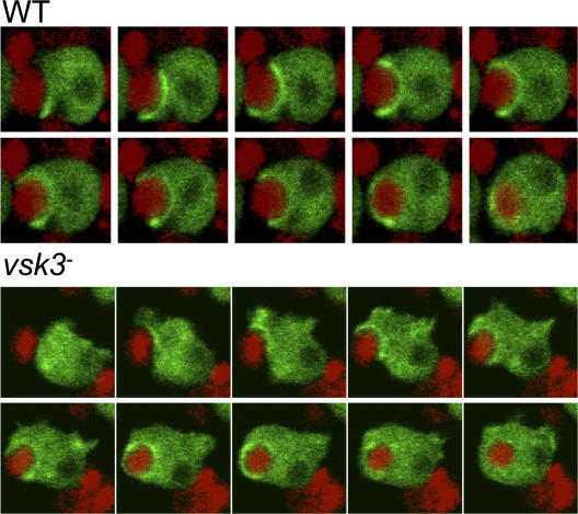
Figure 6.
Dynamic formation of actin-dependent phagocytic cups in wild-type (WT) and vsk3− cells. WT and vsk3 − cells expressing coronin-GFP were fed with TRITC-labeled, heat-killed yeast. Individual cells were imaged simultaneously for GFP and TRITC fluorescence during a time course by confocal microscopy shown left to right at 30-s intervals. See Videos 1 and 2 (available at http://www.jcb.org/cgi/content/full/jcb.200701023/DC1).

