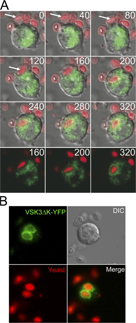
Figure 7.
Late endosomes/lysosomes containing VSK3ΔK-YFP fuse with phagosomes containing newly ingested yeast. (A) Wild-type (WT) cells expressing VSK3ΔK-YFP were fed heat killed, TRITC-labeled yeast and were imaged over time by confocal microscopy to capture the engulfment process, the formation of a phagosome, and phagosome-late endosome/lysosome fusion. The first nine panels show a merged YFP (green), TRITC (red), and DIC time series of a cell ingesting a yeast particle; time in seconds is indicated. The last three frames (time points indicated) are from the same time series without the DIC channel. See Video 3 (available at http://www.jcb.org/cgi/content/full/jcb.200701023/DC1). (B) Higher resolution image of cells in (A) 20 min after feeding showing VSK3ΔK-YFP surrounding yeast filled phagosomes.

