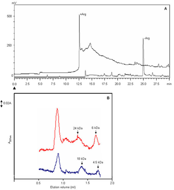
Figure 3. Arginine modulates the chromatographic profile of Aβ1-42.
(A) Reverse phase chromatography of Aβ1-42 peptide. 10 µg of peptide was chromatographed on the RPC C8 column (250×4.6 mm) in the presence and absence arginine. The peptide was eluted with 0–60% acetonitrile linear gradient in PB at a flow rate of 0.7 ml/h and monitored at 257 nm. The arrowhead indicates the start of the gradient. The profiles in the presence and absence of arginine are indicated. (B) Size exclusion chromatography. 10 µg of Aβ1-42 was chromatographed on SMART Superdex G-75 column with and without arginine. The monomeric and tetrameric forms of Aβ1-42 elutes with larger hydrodynamic volume in the presence of arginine (red curve) compared with the control (blue curve). (The molecular weights are indicated by arrows).

