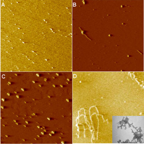
Figure 5. Inhibition of Aβ1-42 fibril formation by arginine and praline.
AFM images (3×3 micron). Aβ1-42 was incubated in PB at 25°C for 24 h. (A) Inhibition of aggregation of Aβ1-42 by 0.2 M arginine. (B) Inhibition of aggregation of Aβ1-42 by 0.2 M proline. (C) Complete solubilization of Aβ1-42 at pH 10.5. No fibrils were observed. (D) Control experiment in which the fibrils formed. (Inset) Transmission electron micrograph of 24 h control sample at higher magnification (22,000×) showing spherical aggregating units.

