
FIG. 2.
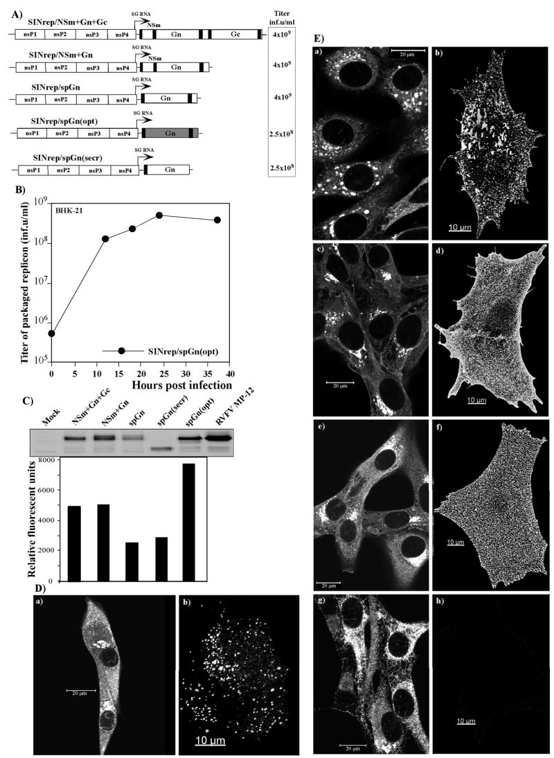
SINV-based replicons used in the study and analysis of the expression of the RVFV-specific glycoproteins by the designed constructs. (A) The schematic representation of the replicons and the titers of the packaged replicons obtained after co-transfection of the replicon and helper RNAs into BHK-21 cells. One of multiple reproducible experiments is presented. The spGn(opt) synthetic gene is indicated in grey color (B) Replication of the tri-component genome virus, having a SINrep/spGn(opt) replicon genome and the genomes of capsid- and glycoprotein-coding helpers, in BHK-21 cells. Cells were infected at an MOI of 8 inf.u/cell, and, at 12 h post infection, the FBS-containing medium was replaced by serum-free VP-SFM medium. Samples of medium were taken at the indicated time points and titers of packaged replicons were determined as described in Materials and Methods. (C) Analysis of RVFV-specific protein expression in cells infected with SINV replicons. BHK-21 cells were infected with packaged replicons at an MOI of 20 inf.u/cells or RVFV MP12 at an MOI of 2.5 PFU/cell and incubated in complete medium at 37°C for 12 h. Equal amounts of cell lysates were analyzed by electrophoresis in SDS-10% polyacryamide gel, followed by Western blotting. Membranes were processed by mouse anti-RVFV antibodies and IRdye 800CW-labeled secondary antibodies. Images were acquired on a Odyssey Infrared Imager (LI-COR). (D) Distribution of virus-specific proteins in the cells infected with RVFV. BHK-21 cells were infected with RVFV MP12 at an MOI of 50 PFU/cell and, at 13 h post infection, stained with mouse anti-RVFV antibodies and AlexaFluor 546-labeled goat anti-mouse secondary antibodies. Panel (a) presents staining of the Triton X-100-permeabilized cells; panel (b) presents surface staining of a non-permeabilized cell. Gn and Gc develop well-distinguishable patches on the cell surface. E) Distribution of the RVFV-specific proteins in the cells infected with packaged SINV replicons that express different fragments of the polyprotein encoded by the M segment of the RVFV genome. Panels (a) and (b) present cells infected with SINrep/NSm+Gn+Gc replicon, panels (c) and (d) present cells infected with SINrep/NSm+Gn replicon, panels (e) and (f) present cells infected with SINrep/spGn, replicon, panels (g) and (h) present cells infected with SINrep/spGn(secr) replicon. Panels (a), (c), (e) and (g) represent images of the cells that were permeabilized with Triton X-100 prior to immunostaining. Panels (b), (d), (f) and (h) present images of the cells stained with antibodies without permeabilization. Images were acquired on a Zeiss LSM510 META confocal microscope using a 63X 1.4NA oil immersion planapochromal lens, as described in Materials and Methods.