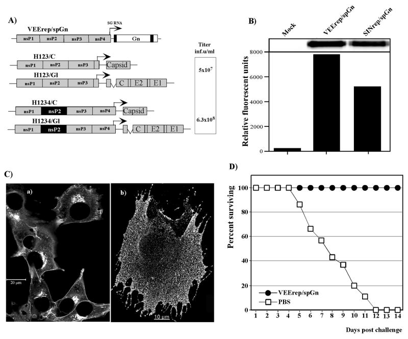
FIG. 3.
VEEV replicon and helpers used in the animal studies. (A) The schematic representation of the RVFV Gn-expressing VEEV replicon and two pairs of helpers encoding VEEV capsid and glycoproteins. Representative titers, obtained in one of multiple, reproducible experiments, are indicated. (B) Analysis of RVFVGn expression in cells infected with SINrep/spGn and VEErep/spGn replicons. BHK-21 cells were infected with packaged replicons at an MOI of 20 inf.u/cells and incubated in complete medium at 37°C for 12 h. Equal amounts of cell lysates were analyzed by SDS-10% polyacryamide gel electrophoresis, followed by Western blotting. Membranes were processed by mouse anti-RVFV antibodies and IRdye 800CW-labeled secondary antibodies. Images were acquired on a Odyssey Infrared Imager (LI-COR). (C) Distribution of RVFV Gn in the cells infected with packaged VEErep/spGn replicon. BHK-21 cells were infected with the packaged replicon at an MOI of 50 inf.u/cell and, at 12 h post infection, stained with mouse anti-RVFV antibodies and AlexaFluor 546 goat anti-mouse secondary antibodies. Panel (a) presents staining of the Triton X-100-permeabilized cells; panel (b) presents cell stained with antibodies without permeabilization. Images were acquired on a Zeiss LSM510 META confocal microscope using a 63X 1.4NA oil immersion planapochromal lens, as described in Materials and Methods. (C) Survival of mice immunized with 5×10⁶ inf.u of packaged VEErep/spGn replicon and challenged in 42 days with 5×10³ PFU s.c. of RVFV ZH501. The control group was injected with PBS and challenged with the same dose of RVFV.

