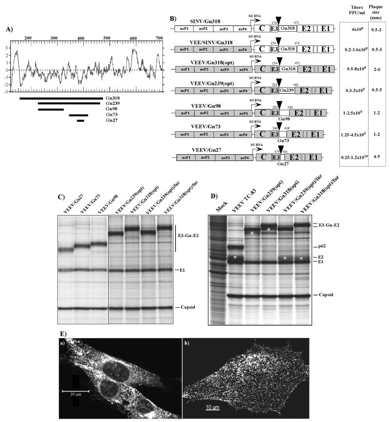
FIG. 6.
The expression cassettes designed for expression of RVFV Gn in viral particles and analysis of the protein expression. (A) The hydrophobicity profile (Kyte-Doolitle) of the Gn protein. Fragments cloned into VEEV structural polyprotein are indicated by black lines. (B) The schematic representation of the recombinant VEEV genomes containing the insertions of the Gn-encoding sequences in the subgenomic RNA. The numbers correspond to the positions of the first and the last a.a. of the insertion in the polyprotein encoded by the RVFV M segment. The triangles indicate positions of engineered furin-specific cleavage sites. VEEV-specific sequences are indicated in grey color. Indicated viral titers and plaque size were derived from multiple experiments. (C and D) The results of metabolic labeling of the proteins expressed by recombinant viruses. The pulse labeling (C) and pulse-chase labeling (D) with [35S]methionine, followed by SDS-10% polyacrylamide gel electrophoresis, were performed at 16 h post infection as described in the Materials and Methods. Final processing products of furin protease are indicated by asterisks. (E) Distribution of Gn-E2 fusion protein in the VEEV/Gn318(opt)-infected cells. Panels (a) represent images of the cells that were permeabilized with Triton X-100 prior to immunostaining with RVFV-specific antibodies. Panels (b) present images of the cells stained with the same antibodies without permeabilization. Images were acquired on a Zeiss LSM510 META confocal microscope using a 63X 1.4NA oil immersion planapochromal lens, as described in Materials and Methods.

