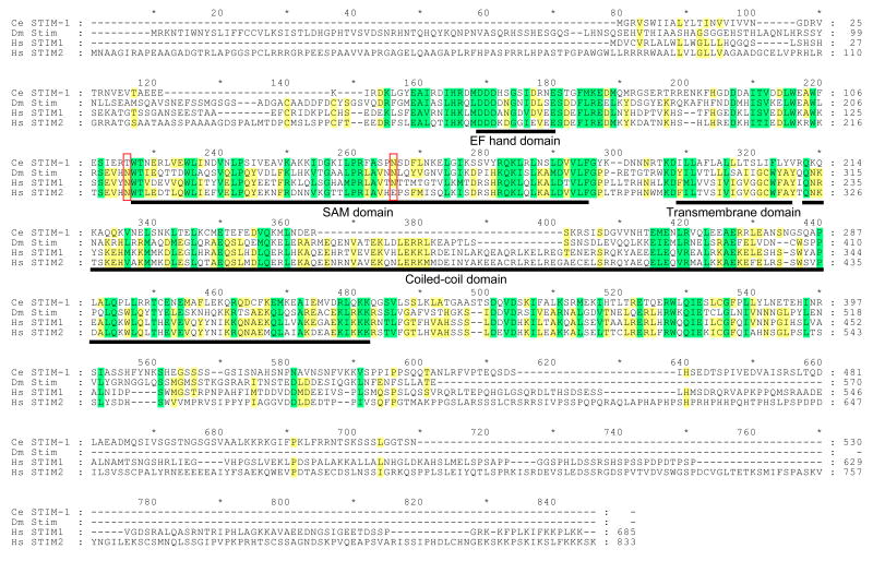
Figure 4.
Amino acid sequence alignment of C. elegans (Ce) STIM-1 with Drosophila (Dm) Stim and human (Hs) STIM1 and STIM2 homologs. Yellow and green shading indicates sequence identity and conserved amino acid substitutions, respectively. Conserved domains are underlined in black. Red boxes show location of N-linked glycosylation sites in human STIM1. Percent similarity and identity of EF-hand, SAM, transmembrane and coiled-coil domains are 58.3% and 50.0%, 50.7% and 37.7%, 27.8% and 5.6%, and 57.4% and 21.3%, respectively. Alignment was performed using Vector NTI software (InforMax, Bethesda, MD). Protein domains were identified by SMART (http://smart.embl-heidelberg.de/smart/change_mode.pl).

