
Figure 2. Expression of CD209 positive dendritic cells in NLGM.
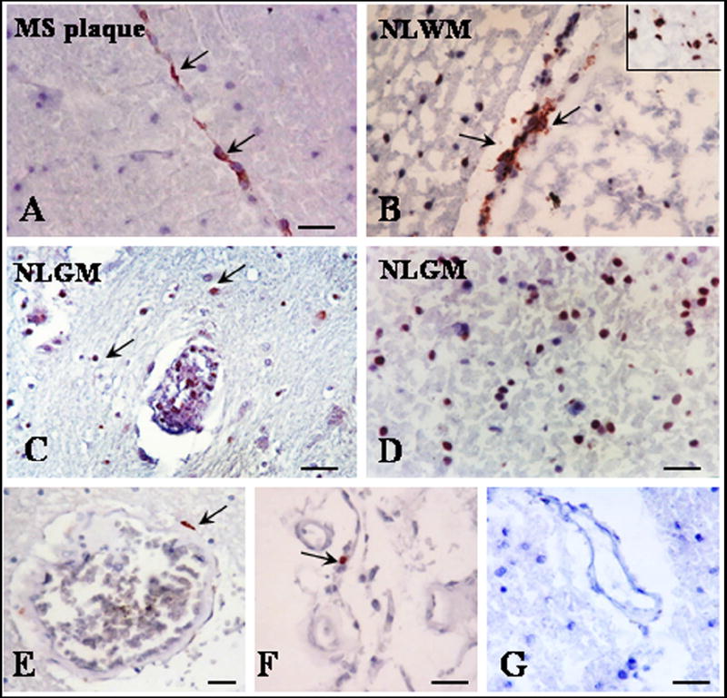
Cryostat sections were stained for CD209 by indirect immunoperoxidase. Perivascular CD209 positive DCs were commonly found in MS plaque (A), NLWM (B) and NLGM (C) (arrows). Parenchymal CD209 deposits were also seen in NLWM (B, insert) and NLGM (D). Rare perivascular CD209 positive cells were seen in control brains white matter (E) and choroid plexus (F). No staining was seen when secondary antibodies were used with non-specific or no primary antibody (G). A-G, Bar= 20 μM. MS plaque= multiple sclerosis plaque, NLGM= non-lesional gray matter, NLWM= non-lesional white matter.