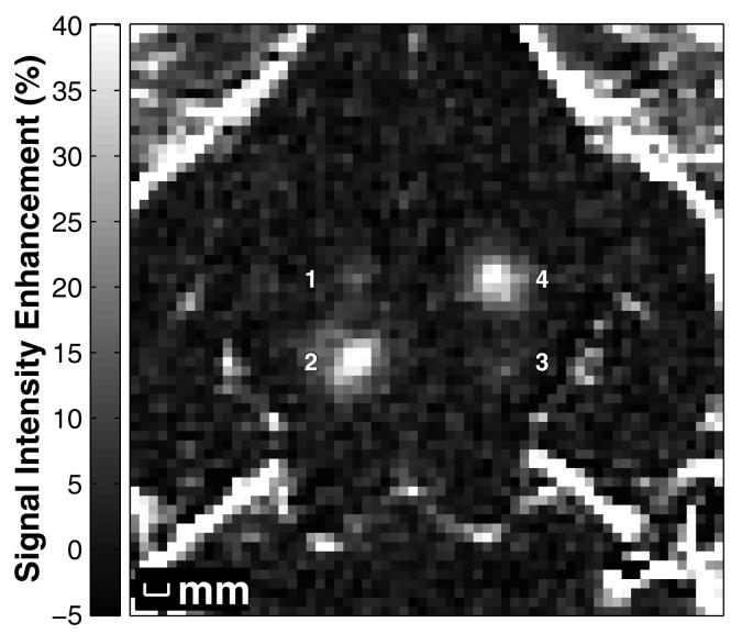
Figure 1.
MR image showing the signal intensity increase due to localized BBBD produced by focused ultrasound pulses combined with Definity® in the rabbit brain at four locations. The signal intensity increase was created from contrast-enhanced T1-weighted images normalized to a T1-weighted image acquired before baseline. The peak rarefactional pressure amplitude (estimated in brain) was 0.2, 1.1, 0.4, and 0.8 MPa for locations 1-4, respectively.

