
Figure 4.
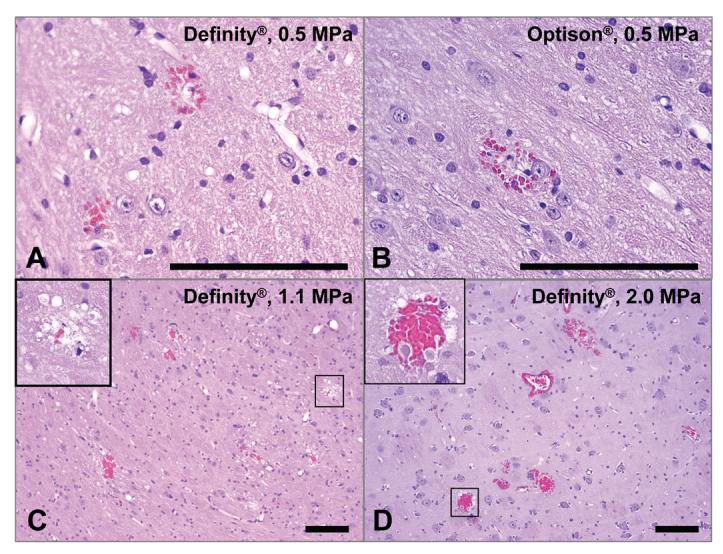
Microphotographs of H&E sections for four sonicated locations with BBBD produced with focused ultrasound combined with Definity® or Optison®. a) – b): histology score = 1; only three areas with extravasated erythrocytes were found in each of these examples. c): histology score = 2; twelve areas with extravasation were found in this example, as well as a vacuolated region (inset). d): histology score = 2; more than 20 regions with extravasations were found in this example, as well as damage (inset). In such cases with more than 20 areas with extravasated erythrocytes edema was evident as well in histology and in MRI. Bar: 100 μm.