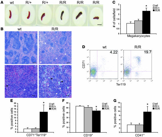
Figure 6. Both erythroid progenitors and megakaryocytes are increased in VhlR/R spleens.
(A) VhlR/R spleens appeared slightly darker in color than WT and VhlR/+ littermates. Scale bar: 1 cm. (B) Representative sections of WT and VhlR/R spleens stained with H&E revealed a disruption of the normal splenic architecture in VhlR/R mice. Erythroid cells are indicated by white arrows, and megakaryocytes by black arrows. Original magnification, ×5 (top); ×40 (bottom). Scale bars: 200 μm; 20 μm. (C) Quantification of the increase in megakaryocytes in VhlR/R mice. *P < 0.006. (D) Representative flow cytometry plots of WT and VhlR/R spleens stained for CD71 and Ter119. (E and F) The percentage of CD71+Ter119+ erythroid progenitors was increased 6-fold (12%) in VhlR/R spleens (E), corresponding to a 9% (1.2-fold) decrease in CD19+ cells (F). *P < 0.05. (G) The percentage of CD41+ cells was also greater in VhlR/R spleens (2.5-fold). *P < 0.05.

