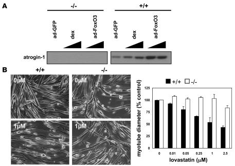
Figure 4. Myotubes from atrogin-1 null (–/–) mice are resistant to lovastatin-induced damage.
(A) Atrogin-1 protein expression is absent in atrogin-1 null (–/–) myotubes. Myoblasts derived from atrogin-1 knockout mice (–/–) and corresponding wild type (+/+) littermates were differentiated into myotubes. Cultures were stimulated to express atrogin-1 with dexamethasone (5 μM) or infected with constitutively active FoxO3- or GFP-expressing adenovirus (45). Atrogin-1 expression was detected by Western blotting as in Figure 3. (B) Myotubes from atrogin-1 null (–/–) and wild-type (+/+) mice were treated with lovastatin at the indicated concentrations for 48 hours. Myotube morphology was examined, and diameter was measured. Original magnification, ×100.

