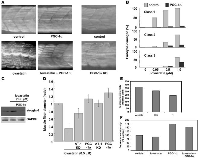
Figure 9. PGC-1α expression reduces lovastatin-induced atrogin-1 expression and muscle damage in zebrafish embryos.
(A) Myosin heavy chain staining of representative zebrafish embryo somites following injection of 100 pg PGC-1α cDNA or vehicle in the presence or absence of 0.5 μM lovastatin for 12 hours (left box) or morpholino oligonucleotides against z–PGC-1α (right box). Note that suppression of atrogin-1 protects from statin-induced damage and that suppression of PGC-1α has a similar muscle phenotype as lovastatin treatment. Original magnification, ×200. (B) Quantitation of muscle damage. Classes of morphological phenotypes are as described in Figure 5. Note that at each lovastatin concentration, the injection of PGC-1α cDNA almost completely abolishes the damage caused by lovastatin. Number of embryos quantitated are 137, 112, 139, and 122 for the controls and 120, 103, 108, and 107 for the PGC-1α–injected embryos at the lovastatin concentrations of 0, 0.05, 0.5, and 1.0 μM, respectively. (C) Western blot of atrogin-1 following 0.5 μM, 12 hours lovastatin treatment in zebrafish embryos injected or not with 100 pg PGC-1α cDNA. (D) Muscle fiber diameter was measured following myosin heavy chain staining as described in Methods in embryos injected with atrogin-1 morpholinos or PGC-1α cDNA. At least 500 fibers were measured at each lovastatin concentration. Results were graphed as the ratio of mean experimental fiber size ± SEM/mean control fiber size ± SEM. Control fiber size: 7.58 ± 0.10 μM. (E) Mitochondrial function is diminished by lovastatin treatment of zebrafish embryos. Cells from zebrafish embryos treated with varying concentrations of lovastatin (0–1 μM) were stained with MitoTracker, and fluorescence intensity, reflecting mitochondrial function, was detected by fluorescence-activated cell sorting. Representative data of mean fluorescence intensity from 3 independent experiments are shown. (F) PGC-1α augments mitochondrial staining and protects against lovastatin’s effects in zebrafish embryos. Embryos were injected with PGC-1α cDNA as described in Methods, then treated with lovastatin (0.5 μM) for 24 hours. As in E, dispersed embryonic cells were stained with MitoTracker and detected by FACS. Data are presented as percentage of mean fluorescence intensity in vehicle-treated embryos.

