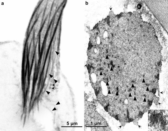
Figure 3.
(a) Confocal view of a longitudinal section cut through the base of a bristle from 36-h-old pupa stained with fluorescent phalloidin. Besides the major cortical bundles are smaller bundles indicated by the arrowheads as well as small round masses (arrows). (b) Transverse section through the base of wild-type bristle from a 36-h pupa. Between some of the cortical actin bundles are surface protuberances in which are snarls of actin filaments (arrows). These are small fluorescent masses indicated by the arrows in a. The arrowheads point to some of the numerous internal bundles. One is shown at higher magnification in the inset.

