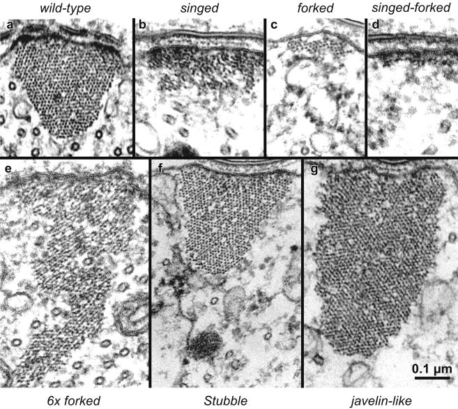
Figure 8.
The concentration of actin filament cross-bridges can determine bundle cross-sectional shape. Representative thin section images of cortical membrane-associated actin bundles in wild-type and mutant microchaetes. All images shown at the same magnification. (a) Wild-type bundles exhibit normal cross-sectional profile in transverse section. Note the triangular shape with a slightly rounded cytoplasmic vertex. (b) The singed mutant bundle forms without the fascin cross-bridge and exhibits a rectangular shape flattened against the plasma membrane. (c) The forked mutant bundle forms without the forked cross-linker and also exhibits a flattened rectangular shape with fewer filaments than singed bundles. (d) The singed-forked double-mutant exhibits an extreme flat, pancake profile composed of a monolayer of filaments attached to the plasma membrane. (e) A bundle formed in the presence of additional forked cross-bridges (6 × forked) contains more filaments than the wild-type and exhibits an extremely “tall” isosceles-like triangle profile that penetrates deep into the bristle shaft cytoplasm. (f) The Stubble mutant exhibits a triangular cross-sectional profile that is larger than wild type. (g) The javelin-like mutant exhibits huge bundles that show rectangular profile in cross-section that extend deeply into the cytoplasm. Of significance is the fact that even though the bundles have very different shapes, the side of the bundle attached to the plasma membrane is approximately the same length in all cases. Bars, 0.1 μm.

