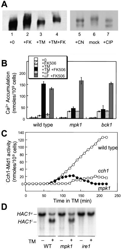
Figure 1.
Identification of kinases required for Cch1-Mid1 Ca2+ channel activation. (A) SDS-urea gradient gel analysis of Cch1-MYC extracted from wild-type cells (strain K1257) treated with tunicamycin (2.5 μg/ml) and FK506 (2.0 μg/ml) as indicated (lanes 1–4). In the right panel (lanes 5–7), Cch1-MYC was first immunoprecipitated from cells grown with tunicamycin and FK506 as described above and treated in vitro with either purified calcineurin, reaction buffer lacking calcineurin, or purified calf intestinal phosphatase. (B) 45Ca2+ accumulation in wild-type cells, mpk1 mutants, and bck1 mutants (strains K1257, RG00993, and RG01328, respectively) was measured after growth for 4 h in SC medium supplemented with tunicamycin, α-factor mating pheromone, and FK506 as indicated. The average of three independent experiments (± SD) is shown. (C) Accumulation of 45Ca2+ in wild-type cells (open circles), cch1 mutants (open squares), and mpk1 mutants (closed circles) (strains K1257, RG04847, and RG00993, respectively) was measured as in A at various times after treatment with tunicamycin plus FK506 and tunicamycin alone. Cch1-dependent Ca2+ influx activity was calculated as the difference between these two measurements at every time point. (D) Northern blot analysis of HAC1 mRNA isolated from wild-type cells, mpk1 mutants, and ire1 mutants (strains K1257, RG00993, and K1259) mutant cells treated with or without tunicamycin for 1 h.

