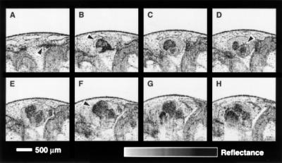
Figure 3.
Sequence of representative in vitro OCT images acquired with the superluminescent diode source illustrating the morphology of the Xenopus heart. High resolution images permit the identification of cardiovascular morphology. (A) Arrow indicates an artery; (B) bifurcation of ventral aorta leaving the truncus arteriosus; (D) atrium; (F) ventricle.

