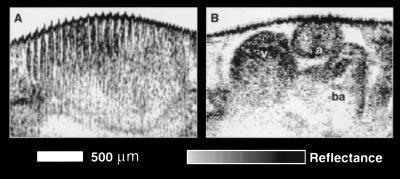
Figure 5.
Comparison of OCT acquisition rates using the (A) superluminescent diode and (B) Cr (4+):forsterite laser. The in vivo Xenopus heart image in A was acquired in 30 s and contains multiple motion artifacts that completely mask the underlying anatomy. Image B was acquired in 0.25 s. The rapid acquisition eliminates the motion artifacts and enables the cardiac morphology to be visualized, in vivo, during the cardiac cycle. a, atrium; v, ventricle; ba, bulbous arteriosus.

