Figure 7.
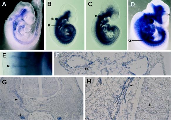
Whole-mount in situ hybridization on mouse embryos using HLF, HIF1α, and flt-1 cRNA probe. (A) Lateral view of 9.5-dpc embryo hybridized with HIF1α cRNA probe. Hybridization signal was seen weakly in the vascular system. ∗, nonspecific signal in the otic vesicle. (B) Lateral view of 9.5-dpc embryo hybridized with HLF cRNA probe. Hybridization signal was observed clearly in the vascular system. F, position of cross-section in F. (C) Lateral view of 9.5-dpc embryo hybridized with flt-1 cRNA probe. (D) Lateral view of 10.5-dpc embryo hybridized with HLF cRNA probe. E, photograph of higher magnification (×45) in E. G and H, position of the cross-section in G and H. (E) Photograph of higher magnification (×45) of dorsal region of 10.5-dpc embryo. Arrows indicate the signals in the intersomitic plexus. (F) Transverse section of 9.5-dpc embryo hybridized with HLF cRNA. Note that HLF mRNA was expressed in the endothelial cells of the dorsal aorta. (G) Transverse section of 10.5-dpc embryo. Arrowheads indicate the signals in the intersomitic plexus. (H) Transverse section of the 10.5-dpc embryo. An arrowhead indicates the signal of the endothelial cells of the sprouting blood vessels. da, dorsal aorta: nt, neural tube: o, optic vesicle; sg, sympathetic ganglion.

