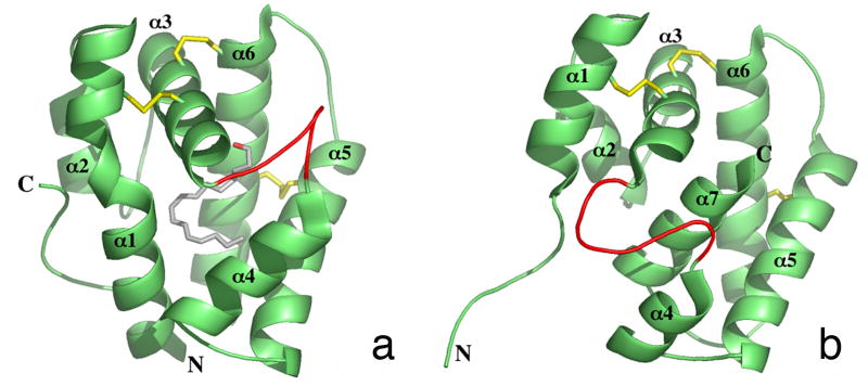
Figure 2. Crystal Structures of Bombykol-Bound and Unliganded BmorPBP.
X-ray crystal structures showing bombykol (A) or no ligand (B) in the binding pocket of BmorPBP (Protein Data Bank ID codes 1DQE and 2FJY, respectively). Secondary structures are depicted in cartoon representation. Bombykol is depicted in ball-and-stick format. Pictures were generated in PyMOL (http://www.pymol.org/).

