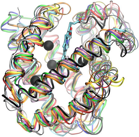
FIGURE 2.
Structure of 10 monomeric globins are aligned and superimposed, demonstrating the very strong conservation of their secondary structure globin fold. The structures are sperm whale (blue), horse (green), and sea hare (cyan) Mbs, soy (black) and lupin (white) Lbs, roundworm (yellow), trematode (red), and bloodworm (orange) Hbs, clam (pink), and midge (purple) Hbs. The Xe binding sites of sperm whale Mb are shown as green spheres, and the proteins' α-helices are displayed as black lines.

