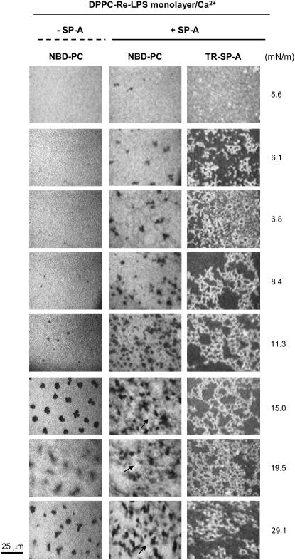
FIGURE 6.
Typical images obtained from a DPPC/Re-LPS (XRe-LPS = 0.2) mixed monolayer containing 1 mol % NBD-PC spread onto a buffered saline subphase containing 150 μM Ca2+, with and without 0.08 μg/ml TR-SP-A at the surface pressures indicated. Images were recorded through filters selecting fluorescence coming either from NBD-PC (emission centered at 520 nm) or TR-SP-A (emission centered at 590 nm). Arrows in the central panel show a distinct, brilliant new phase that dissolves more lipophilic dye. The scale bar is 25 μm.

