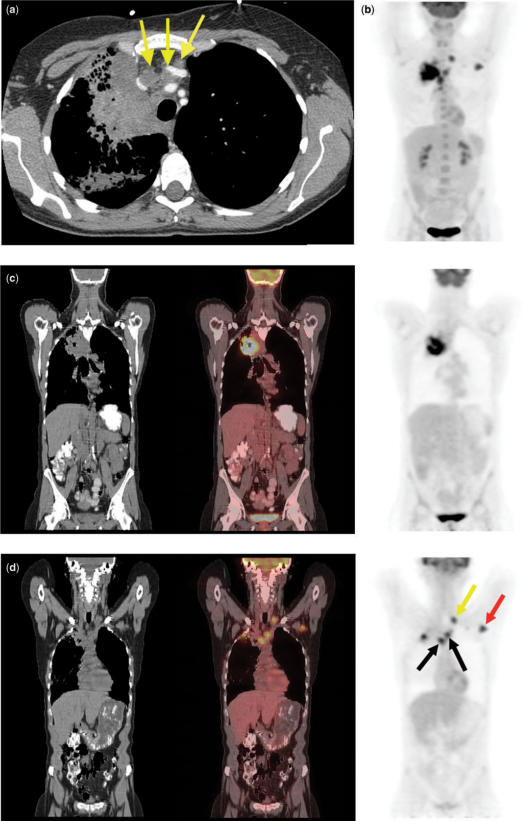
Figure 10.
Right upper lobe squamous cell cancer. Enlarged ipsilateral and contralateral mediastinal lymph nodes at CT (a) (arrows) and bilateral abnormal uptake on a 3D PET image (b) suggest N2 and N3 disease. Integrated coronal CT-PET images (c,d) confirm abnormal uptake within the lung mass (c), as well as bilateral mediastinal lymph nodes (black arrows), a left supraclavicular lymph node (yellow arrow), and a left axillary lymph node (red arrow) (d). Axillary lymph node metastasis represents M1 disease.

