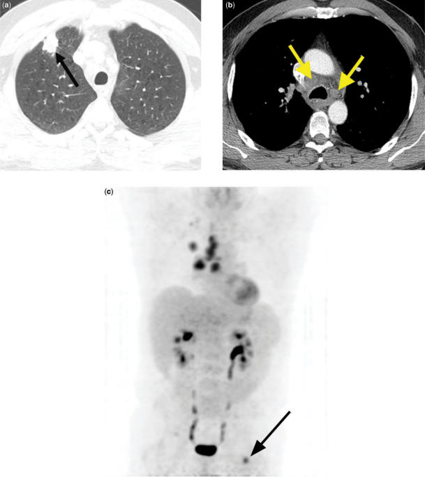
Figure 11.
Right upper lobe adenocarcinoma. CT shows a right upper lobe nodule (a) (arrow) and enlarged ipsilateral and contralateral mediastinal lymph nodes (b) (arrows). 3D PET image (c) confirms abnormal radiotracer uptake within these areas, consistent with N2 and N3 disease. In addition, PET reveals a previously unsuspected distant metastasis in the left femur (M1 disease) (arrow).

