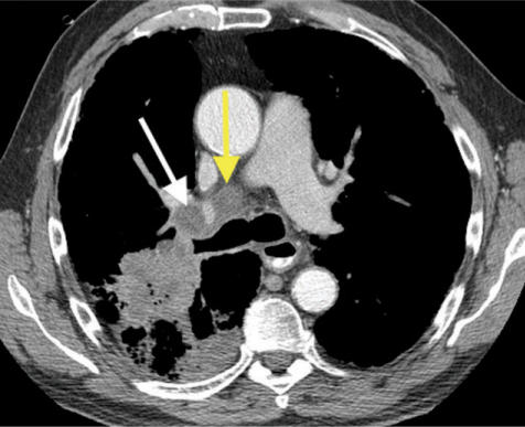
Figure 9.
Right upper lobe squamous cell cancer with post-obstructive pneumonia. CT shows enlarged lymph nodes in the right hilar (white arrow) and tracheobronchial (yellow arrow) regions, suggesting N1 and N2 disease, respectively. Alternatively, these could represent reactive lymph nodes, draining the right upper lobe pneumonia.

