
Fig. 1.

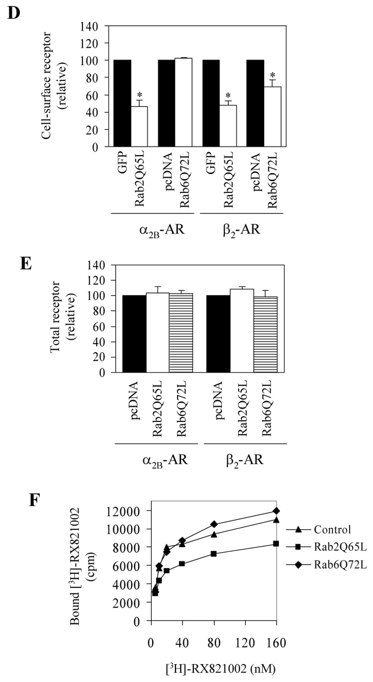
Effect of transient expression of Rab2Q65L and Rab6Q72L on the cell-surface expression of α2B-AR and β2-AR. A. Western blot analysis of GFP-tagged Rab2Q65L expression. HEK293T cells cultured on 6-well plates were transfected with the pEGFP-C1 vector (control) or GFP-Rab2Q65L mutant. Cell homogenates were separated by 12% SDS-PAGE and expression of Rab2Q65L was detected by Western blotting using anti-Rab2 (upper panel) and anti-GFP antibodies (middle panel). B. Western blot analysis of FLAG-tagged Rab6Q72L expression. HEK293T cells cultured on 6-well plates were transfected with the pcDNA3 vector (control) or FLAG-Rab2Q72L mutant and expression of Rab672L was detected by Western blotting using anti-Rab6 (upper panel) and anti-FLAG M2 antibodies (middle panel). β-actin expression is shown in lower panels as a loading control. C. The subcellular distribution of GFP-Rab2Q65L and FLAG-Rab6Q72L. HEK293T cells cultured on coverslips were transfected with GFP-conjugated Rab2Q65L or FLAG-Rab6Q72L. The subcellular distribution of GFP-Rab2Q65L was revealed by fluorescence microscopy detecting GFP and FLAG-Rab6Q72L by immunostaining with anti-FLAG antibodies as described under “Experimental procedures.” Scale bars, 10 µm. D. Inhibition of the cell-surface expression of α2B-AR and β2-AR by Rab2Q65L and Rab6Q72L. HEK293T cells were transfected with GFP-conjugated α2B-AR or β2-AR together with the pEGFP-C1 vector (GFP), GFP-tagged Rab2Q65L, the pcDNA3 vector or FLAG-Rab6Q72L. The expression of α2B-AR or β2-AR at the cell surface was determined by intact cell ligand binding using [³H]-RX821002 and [³H]-CGP12177, respectively, as described in the “Experimental procedures.” The mean values of specific ligand binding were 43371 ± 6368, 44247 ± 3979, 49014 ± 8433 and 51385 ± 2879 cpm (n = 3, each in triplicate) from cells transfected with α2B-AR with pEGFP-C1, α2B-AR with pcDNA3, β2-AR with pEGFP-C1 or β2-AR with pcDNA3, respectively. The data shown are percentages of the mean value obtained from cells transfected with individual receptor and the pcDNA3 or pEGFP-C1 vector and are presented as the mean ± S.E. of three experiments. *, p < 0.05 versus the cells transfected with respective receptor and the pcDNA3 or pEGFP-C1 vector. E. Effect of Rab2Q65L and Rab6Q72L on total expression of α2B-AR and β2-AR. HEK293T cells were transfected with GFP-conjugated α2B-AR or β2-AR together with the pcDNA3 vector, FLAG-Rab6Q72L or Rab2Q65L in the pcDNA3 vector. The overall receptor expression was determined by measuring GFP fluorescence using a flow cytometer as described in the “Experimental procedures.” F. Specific [³H]-RX821002 binding to membrane fractions prepared from the cells transfected with α2B-AR and Rab2Q65L or Rab6Q72L. HEK293T cells were transiently transfected with α2B-AR together with the pcDNA3 vector, FLAG-Rab6Q72L or Rab2Q65L. The membrane preparation (15 µg of protein) was incubated with increasing concentrations of [³H]-RX821002 (5 – 160 nM) for 30 min. Specific binding was determined in duplicate, and nonspecific binding was determined in the presence of 10 µM rauwolscine as described under "Experimental procedures." The data shown are representative of at least three separate experiments, each with similar results.